Blog Credit : Trupti Thakur
Image Courtesy : Google
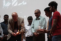
The Portable X-Ray Protection Barrier By IIT Collaboration
Researchers at Sree Chitra Tirunal Institute for Medical Sciences and Technology (SCTIMST) and Government Engineering College, Barton Hill (GECBH) have developed a portable X-ray protection barrier. This device aims to enhance safety during X-ray-based histopathology, a process used to study tissue samples for disease diagnosis.
Collaboration Overview
The project was a collaborative effort between the Biomedical Technology wing of SCTIMST and Hesper Tech Pvt. Ltd., a startup from GECBH’s Technology Business Incubation Centre. M.Tech students from the institutions were instrumental in creating the prototype of this safety device.
How the Device Works
The device is designed to protect healthcare workers from harmful radiation during X-ray examinations of tissue samples. It significantly reduces radiation exposure levels, lowering them from several millihertz to negligible microhertz. This reduction marks a considerable improvement in safety for those conducting histopathology analysis.
Testing and Results
The prototype was tested at the Government Medical College in Thiruvananthapuram. During these tests, a Fluke 451P ion chamber survey meter measured radiation leakage. The results confirmed the device’s effectiveness in minimizing radiation exposure, validating its protective function.
The research team from SCTIMST included Arun Anirudhan, Sabareeswaran A., Ramesh Babu V., and Aneesh K. John. Vimal George and Viswanath S., founders of Hesper Tech Pvt. Ltd., also played crucial roles in the development and testing of the barrier.
About Sree Chitra Tirunal Institute for Medical Sciences and Technology
Sree Chitra Tirunal Institute for Medical Sciences and Technology (SCTIMST), established in 1976, is a premier institution in India specializing in biomedical technology and health research. It operates under the Government of India and is known for its advanced research in fields like cardiovascular devices, diagnostic tools, and rehabilitation technologies. The institute collaborates with various universities, offers Ph.D. programs, and engages in community outreach. With international research partnerships, SCTIMST continues to drive innovation in medical technology and healthcare.
How Patients Are benefitted By this Portable X-Ray Protection
One remote place that has tested ultraportable X-ray hardware is the Orkney Islands in Scotland. Prior to a trial there from November 2021 to January 2022, 73% of patients who were due for an X-ray did not attend their appointments – due in large part to the costs and time needed to reach distant hospitals.
During the trial, a handheld device made by Japanese firm Fujifilm was taken to local clinics. Called the Fujifilm Xair, it weighs just 3.5kg, and is only 301mm (12 inches) wide and 144mm tall.
As a result of its use, the non-attendance rate for X-rays appointments was reduced to zero.
As one patient commented to the subsequent report: “This is hugely beneficial to people on the small islands. Much less upheaval for frailer patients.”
German company OR Technology is another manufacturer of portable X-ray machines. Tim Thurn, its chief commercial officer, says it is seeing great interest from emerging markets. “There’s a huge demand for bringing healthcare to the people,” he says.
In the Philippines, Nigeria and other countries in the developing world, portable X-ray systems are transforming screening for tuberculosis (TB), which kills approximately 1.3 million people a year despite being preventable and curable.
It’s long been known that a chest X-ray is the best screening tool for TB, explains Suvanand Sahu, the deputy executive director of the Stop TB Partnership. This Swiss-based organisation represents more than 1,500 government and non-government bodies around the world.
But he says that access to X-rays was historically hampered by a lack of hospitals. This was particularly the case in remote areas, and among remote, nomadic and displaced groups.
Dr Sahu says that portable X-ray machines, which often include artificial intelligence software to quickly process the images, have been a powerful solution that allows detection to be successfully done out in the field.
“About 10 years ago, if we would have said ‘we can we do an X-ray in the community, with the computer reading it’, I think people would have jumped out of their seats. But it has happened now.”
Dr Sahu adds that the AI has enabled a “quantum leap” in how accurately and quickly these X-rays can be read.
Yet some are worried about the radiation released by portable X-ray machines. Portable X-ray equipment works in the same way as the big, fixed machines in radiology departments in hospitals – the image is created by a targeted burst of ionizing radiation.
In hospitals, x-ray rooms are carefully designed to minimise radiation exposure, for instance with lead walls.
Mr Thurn says that with portable systems this lack of shielding is compensated for by the greater space where they are used, be it outdoors, or in a field hospital setup. In these cases he says that healthcare workers can stand much further away.
However, the issue of radiation needs to be clarified, as currently the international guidelines on radiation safety “are designed for the traditional fixed type of X-ray”, says Zhi Zhen Qin, a digital health specialist at the Stop TB Partnership.
The image quality of portable X-ray machines used to be a concern as well. But Miss Pilkington says that today’s machines rival fixed units in this regard: “The images that are taken on those machines are of a comparable diagnostic standard.”
New Tech Economy is a series exploring how technological innovation is set to shape the new emerging economic landscape.
However, there are barriers to just how small the components of an X-ray system can get. If an X-ray detector is too small to capture a body part in a single exposure, extra images might need to be taken, which would add to time burdens and radiation doses.
And units with a limited battery life or data storage, reduce how many patients can be imaged in one session.
Meanwhile, some systems described as portable aren’t exactly lightweight and easily transported, particularly if they need other hardware to make them work like a stand or computer equipment.
Yet Australian firm Micro-X is now making much lighter ultraportable x-ray machines, due to its new technology that can produce the X-rays without creating heat. This does away with the need for oil and motors for cooling, which add weight.
The high cost of portable X-ray machines is also an issue. While they are cheaper than the larger fixed machines, they are still very expensive.
The Stop TB Partnership says that the price of ultraportable machines ranges between $47,000 and $66,000. There are additional costs for warranties, installation and software.
Dr Suvanand says that as more manufacturers enter this space, “we hope that the competition will drive down the prices”.
Ultimately, his vision for the future is that “everybody who needs an X-ray should have access to this type of modern, digital, ultra-portable X-ray with AI capability”.
Blog By : Trupti Thakur
27
SepThe X-Ray Protection Barrier
Sep 27, 2024Recent Blog
The TechKritiApr 26, 2025

India’s First Quantum Computing VillageApr 24, 2025
India’s Achievement In QKDApr 22, 2025
The V2G TechnologyApr 21, 2025

Country’s Specific Domain By GoogleApr 19, 2025